Frontier Technology Portal Independent technology analysis / Updated daily
Frontier Technology Portal logo
FRONTIER Technology Portal for the next wave of invention

Category: Biotechnology

Synthetic biology, AI drug discovery, diagnostics, genomics, and bio-manufacturing.

  • Biofoundries Automate Experiments, Not Biology Itself

    Biofoundries Automate Experiments, Not Biology Itself

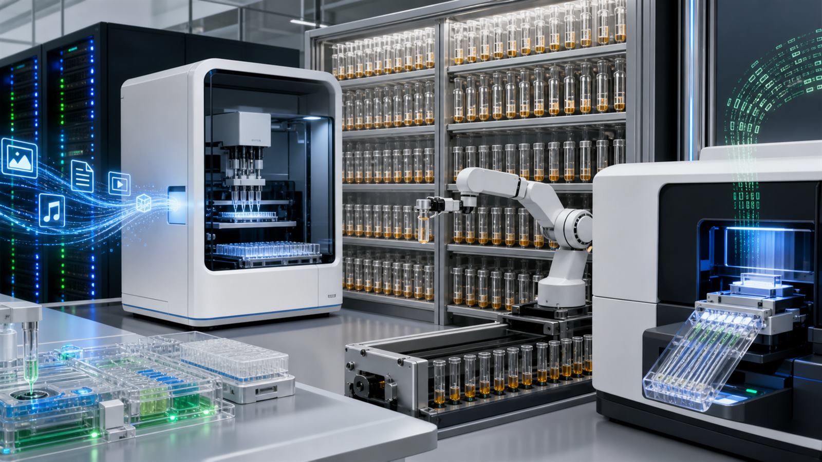
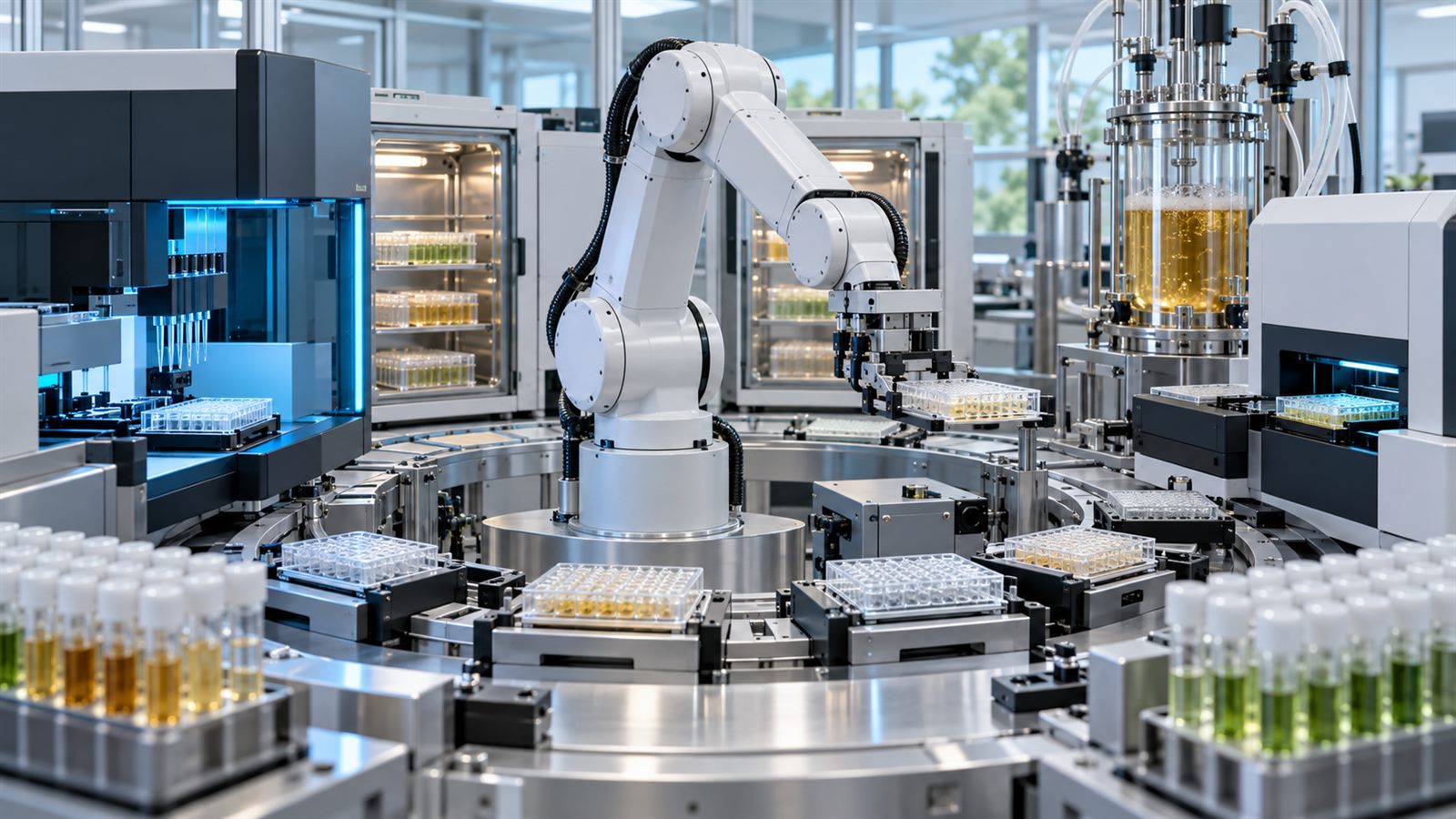
    A modern biofoundry looks less like a traditional biology bench and more like a compact automated factory. Robotic liquid handlers move samples through incubators and analytical instruments. Software schedules experiments, tracks every plate and tube, and feeds measurements into models that propose the next set of designs.

    The goal is not to make biology behave exactly like computer code. Living systems remain variable, context-dependent, and capable of surprising researchers. A biofoundry is valuable because it can run a disciplined design-build-test-learn cycle at a scale and consistency that manual work struggles to match. It automates experiments so that scientists can learn from biology faster.

    A biofoundry is an integrated workflow, not one robot

    Automated pipetting is only one component. A functioning biofoundry connects biological design software, DNA assembly, cell transformation, cultivation, sample preparation, measurement instruments, data storage, analysis, and quality control. Each handoff needs an agreed sample identity, format, volume, timing, and error response.

    The NIST Engineering Biology Program describes this approach as a transition from laborious trial and error toward automated design-build-test-learn, or DBTL. NIST’s Living Measurement Systems Foundry uses automation for microbial growth, manipulation, sample preparation, and measurement while generating data for more predictive engineering.

    Design turns a biological goal into testable variants

    The design stage begins with a target behavior: a microbe that produces a molecule, a protein with improved activity, or a genetic circuit that responds under defined conditions. Software can propose DNA sequences, pathway changes, promoters, protein variants, or experiment combinations. Models help narrow a search space that is far too large to test exhaustively.

    A model’s proposal is still a hypothesis. A DNA part that works in one organism, growth medium, temperature, or genomic location may behave differently in another. Good design therefore includes controls, replicates, and measurements that can distinguish a useful effect from ordinary biological variation.

    This is one reason genome-editing safety depends on sequencing and bioinformatics. A successful edit is not defined only by the intended sequence change; researchers also need evidence about the wider result.

    Build converts digital plans into physical biology

    During the build stage, the foundry assembles DNA, introduces it into cells or prepares cell-free systems, and creates a library of physical variants. Automation can dispense tiny volumes, normalize concentrations, track barcodes, and execute the same protocol across hundreds or thousands of wells.

    That repeatability is useful, but the process is not perfectly deterministic. DNA assembly can fail, cells can grow at different rates, and contamination or evaporation can alter results. The workflow needs checkpoints that confirm identity, purity, and expected construction before later measurements consume time and reagents.

    NIST’s automation program combines liquid handling, incubation, sequencing preparation, cell-free expression, and large experimental designs. The important feature is not that every task is robotic. It is that the tasks are connected to measurement and uncertainty.

    Test needs measurements that are comparable

    The test stage measures what the engineered system actually did. Depending on the project, that may include growth, product yield, protein expression, metabolites, sequence identity, cell fitness, or responses to environmental conditions. High throughput creates more data, but more data is not automatically better evidence.

    Instrument calibration, plate position effects, batch variation, reference materials, and data-processing choices can all change a result. A foundry must record enough metadata to compare experiments across days, instruments, operators, and facilities. Otherwise, automation can reproduce a hidden bias very efficiently.

    The challenge resembles the reproducibility problem for organoid platforms: biological detail is useful only when the workflow can show that measurements are stable and interpretable.

    Learn should choose the next informative experiment

    In the learn stage, statistical models and machine learning connect design choices to measured outcomes. The most useful model may not simply predict the highest performer. It can identify which next experiment will reduce uncertainty or separate competing explanations.

    NIST describes autonomous laboratories as closed feedback loops in which algorithms select samples and measurements to maximize knowledge gained from each iteration. Full autonomy remains an emerging model, and human judgment still defines goals, reviews unexpected results, and decides when evidence is sufficient.

    The loop also depends on negative results. A failed construct or low-producing strain can teach the model where design assumptions break down, but only if the sample history and failure mode are recorded rather than discarded.

    Software and provenance are core laboratory equipment

    A biofoundry needs machine-readable protocols and persistent identifiers for samples, designs, containers, instruments, and data files. The system should be able to reconstruct which source material entered each well, which operations were performed, and which analysis produced a reported result.

    Versioning matters because protocols and software change. If a liquid-handling step, analysis pipeline, or reference database is updated, later results may not be directly comparable with earlier runs. Audit trails make debugging possible and help collaborators reproduce the same process at another site.

    Standards make foundries more than isolated machines

    Different laboratories use different robots, data systems, organisms, and measurement instruments. Common vocabulary, data formats, controls, and performance metrics allow a workflow to travel without requiring identical hardware.

    NIST’s project on engineering biology metrics and technical standards brings together stakeholders from the United States, Europe, Asia, and Australia. It identifies standards and metrics as tools for safety, reproducibility, scale-up, and commerce. The goal is not to freeze innovation, but to make results easier to compare and reuse.

    Scale-up begins after the foundry finds a promising design

    A microbe that performs well in a microplate may behave differently in a large bioreactor. Mixing, oxygen transfer, heat removal, nutrient gradients, foam, contamination control, and downstream purification all change with scale. A biofoundry can optimize a biological design while missing the physical constraints that dominate manufacturing.

    The Agile BioFoundry, a U.S. national-laboratory consortium, links DBTL capabilities to industrial biomanufacturing of fuels and chemicals. Its structure reflects an important point: strain development, process development, and manufacturing translation need to exchange data early rather than operate as separate worlds.

    The same lesson appears in personalized cancer-vaccine manufacturing, where a good molecular design is only one part of a time-sensitive production and quality workflow.

    Automation changes the failure modes

    Robots reduce some manual variation but introduce dependence on scheduling software, deck layouts, calibration, sensors, and data interfaces. A blocked pipette tip or incorrect plate definition can propagate across a large batch before anyone notices. Systems need pause conditions, controls, exception handling, and physical verification.

    Cybersecurity and access control also matter because digital files can encode valuable strains, protocols, and design strategies. Responsible operation requires biosafety review, inventory controls, appropriate screening, and clear authority over what an automated system is allowed to build.

    Limitations and what to watch next

    Biofoundries do not remove the need for skilled biologists, process engineers, or quality specialists. They cannot make a poorly defined objective measurable, guarantee that a model generalizes, or turn a laboratory result directly into a safe commercial product. Not every project needs high throughput; a small number of carefully chosen experiments may be better.

    Watch for more portable protocols, reference materials, shared data models, automated uncertainty tracking, and tighter links between foundry-scale experiments and pilot bioreactors. The decisive advance will not be a robot that runs the most plates. It will be a workflow that produces results other teams can understand, reproduce, and translate into reliable manufacturing.

    Featured image: AI-generated editorial visualization of an automated biofoundry workflow. It is not a photograph of a named laboratory or a hands-on test.
    Primary and authoritative sources

  • A Microbiome Test Is a Measurement Pipeline, Not a List of Bacteria

    A Microbiome Test Is a Measurement Pipeline, Not a List of Bacteria

    A gut microbiome report can make a stool sample look like a census. It may list dozens or hundreds of organisms, assign each a percentage, compare the result with a reference group, and offer a neat diversity score. The precision of the display can hide a harder truth: the report is the output of a long measurement and analysis pipeline.

    What appears in that list depends on how the sample was collected and stored, how cells were broken open, which DNA was amplified, how deeply it was sequenced, which database was searched, and how the software handled ambiguous reads. A microbiome test can be technically informative without proving that a particular organism caused a symptom or that changing it will improve health.

    Microbiota and microbiome are related, not identical

    Microbiota refers to the community of microorganisms in an environment. Microbiome is often used more broadly for those organisms, their collective genetic material, and sometimes the surrounding biological context. Most mail-in tests do not count every living organism directly. They usually extract genetic material from a sample and infer which organisms are represented.

    That distinction matters because DNA can remain after a cell dies, and sequencing does not automatically show what a microbe is doing. A list of genes suggests possible functions. Measuring active RNA, proteins, or metabolites asks different questions. None of these measurements is a complete portrait by itself.

    The sample is a snapshot with a history

    Stool is convenient, but it represents material leaving the intestinal tract rather than a direct map of every location in the gut. Diet, medication, illness, bowel transit time, collection method, temperature, oxygen exposure, shipping delay, and storage can alter what reaches the laboratory or which DNA remains detectable.

    A good protocol records these pre-analytical conditions and keeps them consistent. Negative controls can expose contamination from collection kits or laboratory reagents, while technical replicates can reveal processing variability. The same principle applies beyond human samples. Our guide to wastewater sequencing explains why a composite environmental sample needs carefully defined collection and interpretation boundaries.

    DNA extraction changes the community that becomes visible

    Microbial cells are not equally easy to open. Some have robust cell walls and may require stronger mechanical or chemical disruption. A gentler extraction can recover one group efficiently while underrepresenting another. The amount of human DNA, food material, and other inhibitors in the sample can also affect library preparation and sequencing.

    Extraction is therefore part of the measurement, not a neutral preparation step. When two laboratories use different kits, bead-beating conditions, purification methods, or input masses, their final abundance profiles may differ even if they begin with portions of the same material. A report should identify the method and its quality controls rather than present the result as a method-independent truth.

    16S and shotgun sequencing answer different questions

    16S ribosomal RNA gene sequencing amplifies selected variable regions of a gene found in bacteria and archaea. It is an efficient way to profile broad community structure, but primer choice and the selected region influence what is amplified. Closely related species can share similar sequences, and viruses and fungi are outside the method’s main target.

    Shotgun metagenomic sequencing reads DNA across the sample without targeting only 16S. It can provide greater taxonomic resolution and information about genes that may be present, including material from organisms missed by a bacterial 16S survey. It also requires more sequencing and computation, can be affected by host DNA, and still depends on reference databases. Our article on long-read sequencing describes the broader reason read length and reference quality affect what genomic analysis can resolve.

    The National Human Genome Research Institute’s Human Microbiome Project overview describes how researchers used both 16S and metagenomic sequencing. The methods are complementary, so a consumer should ask which one produced a report before comparing it with another service.

    Relative abundance is not an organism count

    Many reports show relative abundance: the fraction of classified reads assigned to each taxon. Because the fractions must add to 100 percent, one organism’s percentage can rise when another falls even if its absolute cell count does not change. DNA extraction efficiency and the number of gene copies per organism can further separate read share from cell share.

    A diversity score compresses the number and distribution of detected taxa into one value. It may be useful for comparing samples processed by the same method, but it is not a universal health grade. Different body sites and biological contexts can have different appropriate community structures, and two communities with the same diversity score can contain very different organisms.

    Software and databases make interpretive choices

    After sequencing, software removes low-quality reads, separates human from microbial material, assigns sequences to taxa, and calculates abundance. Results depend on alignment rules, confidence thresholds, database version, naming conventions, and how the pipeline treats organisms with nearly identical sequences.

    A database cannot identify a genome it does not represent well. Classification can also change as reference genomes are corrected or taxonomic names are revised. Predicting metabolic pathways from detected genes adds another layer of inference: a gene may be present without being active, and similar sequences do not always produce identical behavior.

    Reference material reveals pipeline bias

    In 2025, the US National Institute of Standards and Technology released Human Fecal Material RM 8048. It contains stable, homogeneous pools characterized for microbial species and metabolites. Laboratories can process the same reference material and compare their results with NIST’s measurements and with other laboratories.

    This does not make stool simple. It makes variation visible. If a pipeline consistently misses a known component, overstates another, or changes after a software update, the reference material can expose that shift. NIST’s microbiome measurement program emphasizes that standards are needed to benchmark metagenomics and metabolomics before research findings can translate reliably into diagnostics, therapeutics, environmental monitoring, or biosurveillance.

    Reference material tests analytical performance. It does not prove clinical meaning. A method can accurately detect a microbial pattern while the connection between that pattern and a disease, treatment response, or useful action remains uncertain.

    Analytical validity is not clinical utility

    Analytical validity asks whether the test reliably measures what it claims to measure. Clinical validity asks whether that measurement is associated with a health condition in the intended population. Clinical utility asks whether using the result improves decisions or outcomes. These are separate evidence requirements.

    The US Food and Drug Administration notes in its direct-to-consumer testing guidance that evidence and regulatory review vary by test and intended use. A wellness report, a research assay, and a medical diagnostic claim should not be treated as interchangeable. Consumers should check the exact claim and authorization status instead of assuming that every laboratory-generated report has independent clinical validation.

    How to read a report without overreading it

    Start with the method: sample type, collection conditions, extraction protocol, sequencing approach, database version, and quality controls. Look for a clear reference population and ask whether it matches the report’s intended use. Treat taxonomic percentages as method-dependent estimates, not exact cell counts.

    Be cautious when a report turns association into causation or recommends supplements, diets, or treatment from one sample without validated evidence. Microbiome profiles can change, so an unexplained difference between two dates may reflect biology, sampling, or pipeline variation. Health decisions should be discussed with an appropriately qualified clinician rather than based on this article or a general consumer report.

    Limitations and what to watch next

    Even well-controlled sequencing cannot capture every organism, strain, metabolite, or interaction in the gut. Reference materials represent selected samples, not the full diversity of people, diets, geographies, ages, and health states. Privacy also matters because a metagenomic file can contain human DNA as well as microbial sequences.

    Watch for wider use of shared reference materials, interlaboratory proficiency testing, transparent pipeline versioning, absolute-abundance measurements, and prospective studies that connect a predefined test with a useful clinical outcome. The most credible microbiome products will explain where uncertainty enters the pipeline and separate a reproducible measurement from a health claim.

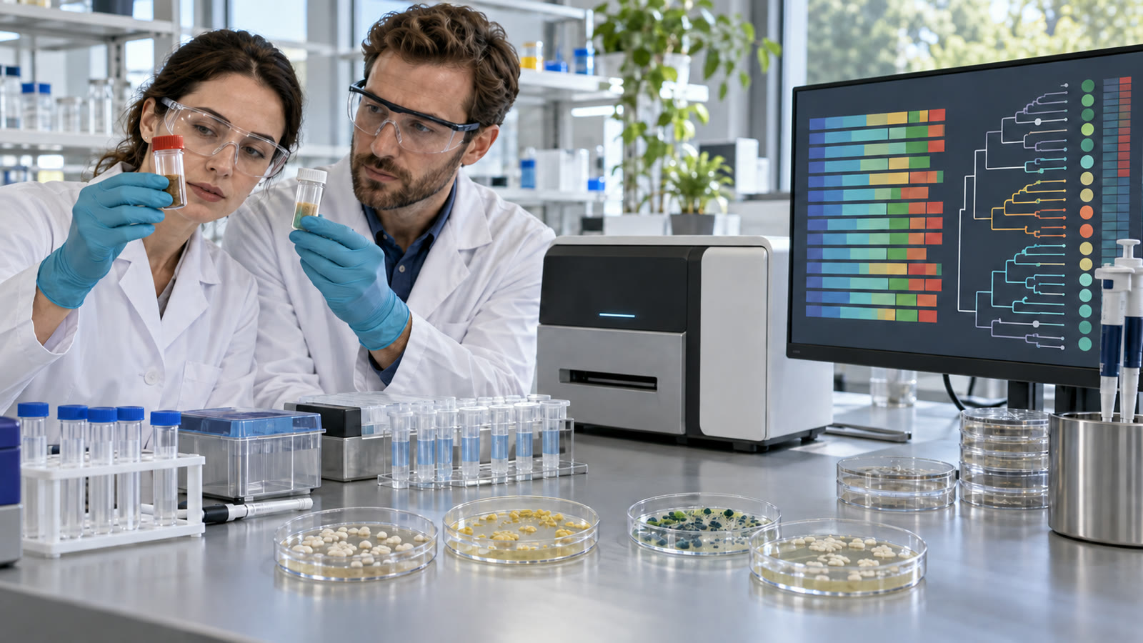
    Featured image: AI-generated editorial illustration of a microbiome measurement workflow, not a photograph of a specific laboratory or diagnostic product.

    Primary and authoritative sources

  • Organ Chips Need Validation, Not Just More Biological Detail

    Organ Chips Need Validation, Not Just More Biological Detail

    Organ-on-chip systems look deceptively simple: a transparent device, tiny channels, flowing liquid, and living human cells. Yet the goal is ambitious. Researchers want those cells to recreate a useful part of an organ’s behavior well enough to predict how a drug, chemical, infection, or disease process will behave in people.

    That promise is why tissue chips are attracting attention in drug discovery and safety research. The technology can add human-relevant biology between a flat cell culture and a clinical trial. But a chip that looks biologically impressive is not automatically ready to guide a high-stakes decision. The decisive question is validation: does it produce repeatable, relevant results for a clearly defined use?

    A chip is a model, not a miniature person

    The US National Center for Advancing Translational Sciences describes tissue chips as bioengineered devices that use human cells to mimic parts of organs such as the heart, kidney, and lung. A chip may place different cell types on opposite sides of a porous membrane, expose them to flowing media, or apply mechanical stretching.

    Those features can reproduce an interface that a conventional dish cannot: air and blood in a lung model, filtration in a kidney model, or transport across a barrier. They do not recreate every cell type, immune signal, hormone cycle, nerve input, and long-term adaptation of a living body. The value of a model comes from being fit for a purpose, not from claiming to be a complete organ.

    Context of use comes before complexity

    A liver chip might be useful for an early toxicity screen, a study of a transport mechanism, or comparing a short list of compounds. Each use needs different evidence. A model that reliably detects one known injury mechanism may not predict every form of human liver toxicity.

    NIH’s organ-on-chip initiative notes that researchers are working across many organ systems, while also recognizing that results must be validated before these models can replace established approaches. That distinction is healthy. A sophisticated platform with flowing channels is not automatically more informative than a simpler assay for every question.

    Before adopting a chip, teams should write down the decision it will support, the endpoint it must measure, the acceptable false-positive and false-negative rates, and the reference data against which it will be judged.

    Reproducibility is an engineering problem

    Living systems vary. Donor cells differ by genetics, age, health history, and source. Cells can change with passage number and culture conditions. Flow rate, temperature, oxygen, media composition, chip material, and the timing of a dose can all affect an outcome.

    That does not make organ chips unusable. It means the platform needs controlled inputs, documented protocols, predefined acceptance criteria, and performance checks. A useful report should identify cell source and characterization, device materials, flow conditions, readouts, controls, and excluded runs.

    The NIH Standardized Organoid Modeling Center is explicitly focused on reproducible, reliable, and accessible models. Standardization is often less glamorous than a new chip design, but it is what lets a result travel between laboratories.

    Human cells can improve relevance without eliminating uncertainty

    Human cells may reveal pathways that differ from animal models or immortalized cell lines. That is particularly valuable when a drug’s target, metabolism, or toxicity is species-specific. Tissue chips can also make it easier to observe human barrier tissues and multi-cell interactions under controlled conditions.

    However, a human-cell system can still be incomplete, immature, or stressed by the culture environment. A model built from a narrow donor pool may not represent the patients a medicine is intended to serve. Researchers must avoid replacing one oversimplification with another.

    The same discipline applies to other emerging measurement systems. Our article on optical clocks explains why a remarkable instrument still needs agreed comparisons. In biology, agreed reference compounds and shared performance metrics play a similar role.

    Multi-organ links add realism and new failure modes

    Connecting liver, gut, kidney, or heart modules can model how a compound is absorbed, transformed, and cleared across tissues. NCATS describes tissue chips as modular systems that can be connected to examine effects on several organ systems.

    But each connection introduces a new challenge. Different tissues may need different media, flow rates, oxygen levels, and time scales. A shared circulation loop can dilute signals or introduce material absorption effects. A failure in one module can be misread as a biological interaction.

    For many decisions, a single well-characterized tissue may be more useful than a complicated multi-organ system. Complexity should earn its place by improving prediction for the stated question.

    Where organ chips fit in drug development

    Organ chips can help prioritize candidates, investigate a mechanism, compare formulations, or flag a possible safety signal. They can reduce the number of unpromising compounds that move into later studies. They may also complement animal studies and clinical evidence where human-specific biology matters.

    They do not erase the need for careful clinical trials. A chip cannot establish population-level safety, rare side effects, long-term outcomes, or real-world adherence. It produces evidence at a specific biological scale.

    Good data management matters as much as the device. Provenance of cells, protocols, images, and analysis code should be traceable, echoing the value of build provenance in software systems. Traceability makes it possible to investigate why one run differs from another.

    What to ask when evaluating a claim

    Ask what outcome the chip predicts, which reference data were used, how many independent runs and cell donors were tested, whether blinded comparisons were performed, and how the model performs against existing methods. Check whether the endpoint is relevant to the intended decision rather than merely visually striking.

    Also ask about throughput, cost, automation, data analysis, and failure handling. A platform can be biologically strong but too slow or variable for routine screening. Conversely, a fast platform may be most useful as an early filter rather than a final decision tool.

    Limitations and what to watch next

    Key limitations include incomplete tissue maturity, donor variability, missing systemic biology, material interactions, and uneven standards across laboratories. The next advances will likely come from shared reference compounds, standardized readouts, representative cell sources, automated manufacturing, and evidence that a model improves a real development decision.

    Organ chips are not a shortcut around biology. They are a promising way to ask more human-relevant questions earlier. Their credibility will depend on reproducibility and transparent validation, not on how closely a device resembles a tiny organ in a photograph.

    Primary and authoritative sources

  • DNA Data Storage Needs a File System, Not Just Dense Molecules

    DNA Data Storage Needs a File System, Not Just Dense Molecules

    DNA is often introduced as an almost impossibly dense storage material. In principle, a small quantity of synthetic DNA can represent a large digital archive, and dry DNA can remain stable far longer than magnetic tape when it is prepared and stored correctly. Those properties make it attractive for data that must survive for decades but is rarely opened.

    Density and durability, however, are only properties of the medium. A usable archive also needs a file format, addresses, error correction, a way to find one object without reading everything, durable packaging, and enough metadata for a future system to decode the molecules. DNA storage will become practical only when that complete chain is reliable and affordable.

    DNA is the medium, not the file system

    Ordinary storage hides several layers. An application creates a file, a file system assigns locations and metadata, a controller protects blocks against errors, and a drive turns those blocks into physical states. DNA storage needs equivalents for all of them, even though its physical medium is a pool of molecules rather than ordered sectors on a disk.

    The SNIA DNA Data Storage Technology Review, published in 2025, describes an end-to-end pipeline that includes coding, DNA synthesis, preservation, retrieval, sequencing, and decoding. Demonstrations have connected those stages, but the review characterizes commercialization as nascent. The important comparison is therefore not DNA density against a hard drive platter. It is one complete, recoverable archive against another.

    A codec translates bits into constrained molecules

    A DNA storage codec maps binary data into sequences built from adenine, cytosine, guanine, and thymine. A naive two-bits-per-base mapping looks efficient, but synthesis and sequencing do not treat every possible sequence equally. Long runs of one base, extreme ratios of certain bases, and repeated patterns can raise error rates or complicate identification.

    Practical encoders add sequence constraints, indexes, checks, and redundancy. They may divide a file into many short packets because current systems generally synthesize short strands rather than one chromosome-length data object. Every extra address or parity symbol reduces headline density, but removing those protections can make the archive impossible to reconstruct.

    This is a familiar lesson from biotechnology. Our explanation of programming cells with synthetic biology shows that DNA is a physical substrate with context-dependent behavior, not an abstract string that can be manipulated without constraints.

    Errors are more complicated than a flipped bit

    Electronic storage often models an error as a bit changing value. DNA pipelines can introduce substitutions, insertions, deletions, missing strands, uneven numbers of copies, and contamination. Some molecules may never be recovered from a sample, while others appear many times in the sequencing output.

    Error-correcting codes and consensus decoding compensate for these effects. Addresses help group reads that belong to the same packet, and redundancy lets software reconstruct missing information. The decoder also has to decide which reads are genuine enough to use. Long-read and short-read instruments have different error profiles; our guide to long-read sequencing and short-read limits explains why read length alone does not determine data quality.

    A storage claim should therefore report the original data size, synthesized bases, redundancy, sequencing coverage, failed strands, recovered bytes, and final error rate. Quoting only theoretical bases per gram conceals most of the engineering problem.

    Random access must avoid reading the entire pool

    An archive containing millions of short strands is physically unordered. To retrieve one file, a system needs molecular addresses and a selective operation that enriches the desired strands before sequencing. Otherwise, opening one photograph could require reading the whole archive.

    A landmark 2018 Nature Biotechnology experiment stored more than 200 megabytes across over 13 million DNA oligonucleotides. The researchers used a primer library to select individual files and reported error-free recovery of 35 files. This showed that large-pool random access is possible, but it did not remove the need to manage primer interactions, amplification bias, sample handling, and repeated access.

    Researchers continue to explore alternatives. A 2025 Nature Communications study used CRISPR-Cas9-based selection and machine-guided sequence design to support random access and content-oriented search. It is promising research, not evidence that commercial DNA archives can already search arbitrary content at cloud-storage speed.

    A future reader needs instructions from the archive itself

    A durable medium is not useful if the decoder disappears. A DNA vial stored for a century may outlive the company, software, sequencer model, and codec that created it. The archive needs a discoverable bootstrap record describing its format, organization, codec, and required interpretation steps.

    SNIA’s DNA Data Storage Sector Zero specification addresses this problem by defining a recommended method for storing vendor and codec information needed to interpret the rest of an archive. It is the molecular equivalent of leaving a readable map at the entrance, although the map still depends on agreed conventions and a compatible reader.

    Interoperability will require more than one specification. Archive identity, containers, indexes, error metrics, environmental records, and migration procedures all need stable definitions. Open test archives will be important because two implementations can follow the same broad idea while making incompatible assumptions.

    The physical container is part of the storage system

    DNA stability depends on moisture, heat, oxygen, light, chemical preparation, and packaging. Dry, protected samples can be durable, but a laboratory tube is not automatically a century-scale product. Designers need validated containers, environmental limits, tamper evidence, inventory controls, and procedures that prevent one retrieval from degrading the remaining material.

    Synthetic data DNA is not intended to function as a living organism, yet sequence screening and physical containment still matter. Encoders should avoid creating sequences with unintended biological significance, and facilities need controls against sample mix-ups or unauthorized copying. Encryption remains necessary for confidential information because anyone who obtains and sequences an unencrypted sample may be able to reconstruct it.

    Writing cost and latency define the realistic market

    DNA synthesis is the write operation, and sequencing is the read operation. Both involve biochemical processing, instruments, consumables, and time. Performance is measured in hours or longer for many workflows, not the microseconds expected from memory or solid-state storage. Rewriting a small block is also awkward when the archive consists of sealed molecular packets.

    That points toward a cold archival tier: information written once, stored compactly with little operating energy, and read rarely. Scientific records, cultural preservation, and mandated retention are more plausible early uses than active databases, video streaming, or backups that must be restored immediately. Tape remains a formidable competitor because it has standardized formats, mature robotics, known costs, and a large installed base.

    How to evaluate a DNA storage claim

    Ask whether the result covered the full write-store-retrieve-read cycle or only one stage. Check net user data after indexes and redundancy, recovery rate across independent samples, random-access selectivity, storage conditions, time to first byte, total throughput, energy, cost, and whether the archive was opened more than once.

    Also look for an escape path from the original vendor. A serious archive should explain how a future reader discovers the codec, verifies integrity, replaces obsolete equipment, and migrates data without losing provenance. Demonstrating that DNA can hold bits is no longer enough; demonstrating operational custody over decades is the real test.

    What to watch next

    The most meaningful progress will be cheaper high-throughput synthesis, less destructive random access, standardized archive metadata, independent interoperability tests, and long-duration stability evidence. Better codecs will matter, but so will robotics, packaging, sequencing economics, and auditable chain of custody.

    DNA storage is credible as a future archival medium precisely because its limitations are becoming clearer. The winning system will not be the one with the most dramatic density calculation. It will be the one that can reliably return the right file, prove that the bytes are intact, and tell a future reader how to do it again.

    Primary and authoritative sources

  • Long-Read Sequencing Exposes Structural Variation Short Reads Can Miss

    Long-Read Sequencing Exposes Structural Variation Short Reads Can Miss

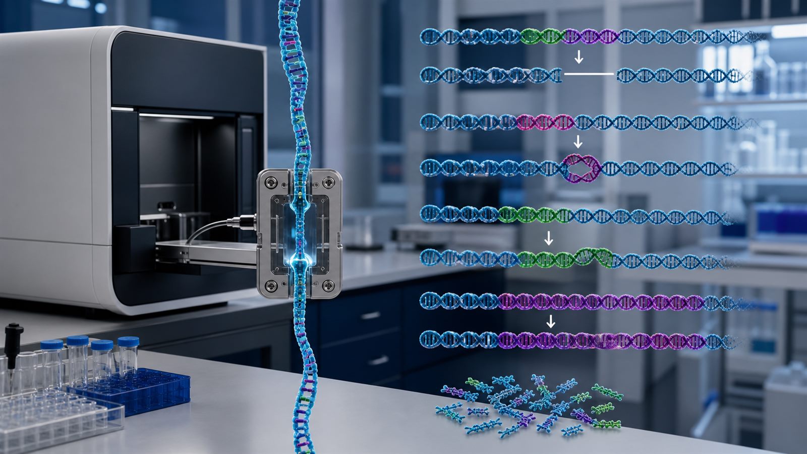
    A human genome is not only a three-billion-letter string dotted with single-letter changes. Large segments can be deleted, duplicated, inverted, inserted, moved, or repeated a different number of times. These structural variants can reshape genes and their regulation, yet many are difficult to reconstruct from conventional sequencing that reads DNA in short fragments.

    Long-read sequencing approaches the same genome with molecules that may span thousands to hundreds of thousands of DNA bases. A single read can cross a repetitive region and connect the unique sequence on both sides. That extra context can turn an ambiguous pile of fragments into a recognizable event. It does not make genome interpretation automatic, but it changes which variants scientists can observe with confidence.

    Structural variation changes more than one DNA letter

    Structural variant is a broad term commonly applied to changes of roughly 50 bases or more. A deletion removes a segment; a duplication adds another copy; an inversion reverses a region; an insertion adds sequence that is absent from a reference; and a translocation joins material from different locations. Tandem repeats can expand or contract, sometimes by many repeat units.

    The boundaries matter. A rearrangement can interrupt a protein-coding gene, alter the number of gene copies, move an enhancer, or change the three-dimensional neighborhood that controls gene activity. Some variants have clear biological consequences, many are harmless, and others remain uncertain. Detection is therefore the beginning of analysis, not a diagnosis.

    The National Human Genome Research Institute explains that long-read methods are especially useful for repetitive and complex genomic regions because their reads extend much farther than typical short-read sequences. That ability is the central advantage, even though different instruments use different sensing chemistry and analysis pipelines.

    Short reads lose context inside repeated sequence

    Imagine tearing several copies of a nearly identical paragraph into tiny strips and then trying to rebuild the original pages. A strip containing a common phrase may fit in several places. DNA repeats create the same mapping problem. If a read is shorter than the repeated region, software may not know which copy produced it.

    Paired short reads can provide distance and orientation clues, while changes in coverage can reveal gains or losses. Sophisticated callers combine those signals with split alignments and local assembly. They detect many important variants, especially in well-characterized regions. Problems grow when breakpoints lie inside long repeats, when inserted sequence is not represented in the reference, or when several changes occur close together.

    A long molecule can anchor in unique sequence before the repeat, travel through the difficult section, and anchor again afterward. That direct continuity helps place breakpoints, measure repeat length, and identify the inserted sequence itself. It can also distinguish changes on the chromosome inherited from one parent from those on the other.

    Long reads support phasing and genome assembly

    Humans normally carry two copies of each autosomal chromosome. Standard reference-based analysis can list variants without showing which changes occur together on the same copy. Long reads often contain multiple nearby markers, allowing software to phase them into longer haplotypes. This can reveal whether two disruptive variants affect one gene copy or both.

    Long reads also improve de novo assembly, in which a person’s sequence is reconstructed into large contiguous pieces before comparison. Assembly-based analysis can represent complex alternatives that are awkward to describe only as edits against one linear reference. It is particularly useful in duplicated regions, immune-related loci, and areas near centromeres and other repeat-rich structures.

    Large population projects are showing the scale of this difference. A 2025 Nature study of 1,019 individuals used long-read sequencing to characterize structural variation across diverse human genomes. Another 2025 study produced near-complete assemblies from 65 human genomes, expanding the set of sequence and structural variation available for population analysis.

    Better detection does not remove technical error

    Long-read platforms have improved substantially, but accuracy depends on the technology, chemistry, run conditions, base-calling model, coverage, and genomic context. Some workflows generate highly accurate consensus reads from repeated observations of the same molecule. Others emphasize very long molecules and real-time output. The right choice depends on whether a project prioritizes single-base accuracy, maximum length, methylation signals, speed, or cost.

    DNA extraction is a practical constraint. Long molecules break under rough handling, and archived or formalin-fixed tissue may already be fragmented or chemically damaged. High-molecular-weight preparation can require more careful collection and quality control than a short-read workflow. A nominally long-read instrument cannot restore information that was lost before the sample reached it.

    Coverage also matters. Low coverage can miss a variant or support the wrong assembly path, especially in a mosaic sample where only a fraction of cells carry the change. Repeats longer than the reads can remain unresolved. GC-rich sequence, homologous genes, and extremely large rearrangements can still challenge both sequencing and software.

    Benchmarks are essential for knowing what a pipeline misses

    A variant caller can produce a polished file without revealing every blind spot. Developers need reference materials with carefully characterized variants so they can measure precision, recall, breakpoint accuracy, and performance by genomic region. Comparisons should separate easy sequence from repeats and other difficult contexts instead of reporting one reassuring average.

    The US National Institute of Standards and Technology coordinates the Genome in a Bottle consortium, which develops benchmark genomes, data, and methods for evaluating sequencing results. Its HG002 benchmark work includes small variants and structural variants. Such resources help laboratories validate an entire workflow, including sample preparation, sequencing, alignment or assembly, variant calling, filtering, and reporting.

    Independent confirmation remains appropriate for findings with major clinical or research consequences. The validation method should match the event. A small breakpoint assay may confirm a junction but fail to establish the full size or copy number of a complex rearrangement. Orthogonal evidence can include targeted sequencing, optical mapping, cytogenetics, copy-number assays, or family data.

    A variant call is not the same as an explanation

    Finding more variants increases the interpretation workload. Population frequency, inheritance, gene function, dosage sensitivity, regulatory context, and phenotype all influence whether a change is meaningful. A rare structural variant is not automatically harmful, and absence from a database may reflect limited sampling rather than danger.

    This distinction parallels our discussion of sequencing in genome-editing safety analysis: a measurement method can strengthen detection without deciding clinical significance on its own. For individualized treatment, as in the case of personalized CRISPR platforms for rare disease, the chain from molecular evidence to intervention requires separate functional, manufacturing, and regulatory evidence.

    Long reads can connect sequence with other molecular layers

    Some long-read technologies can infer base modifications from the same physical signal used to read DNA. That creates an opportunity to associate structural variation, haplotype, and methylation without splitting the sample into entirely separate assays. Long RNA reads can similarly capture full transcript isoforms and show how rearrangements alter splicing.

    Those capabilities complement, rather than replace, methods that preserve location in tissue. Our overview of spatial proteomics explains why molecular identity and tissue context answer different questions. A resolved genome can identify a rearrangement, while spatial and cellular measurements may be needed to see where its downstream effects occur.

    What ordinary readers should watch next

    The important progress is not simply a higher maximum read length. Watch for better accuracy in medically relevant repeats, more representative population references, transparent performance by variant class, interoperable pangenome tools, and benchmarked end-to-end workflows. Falling cost and simpler sample preparation will determine whether long reads move from specialist centers into routine laboratories.

    Researchers should also report what remains inaccessible. A study’s coverage, molecule-length distribution, benchmark regions, confirmation strategy, ancestry mix, and sample quality can matter as much as its headline variant count. Stronger instruments reveal more of the genome, but responsible conclusions still depend on calibrated uncertainty.

    Long-read sequencing is valuable because genomes are physical molecules, not disordered bags of letters. Preserving more of each molecule retains the context needed to cross repeats, phase chromosomes, and reconstruct complex events. The result is a more complete map of structural variation, paired with a new obligation to validate and interpret what that map contains.

    Primary and authoritative sources

  • Wastewater Sequencing Tracks Community Pathogens, Not Individual Cases

    Wastewater Sequencing Tracks Community Pathogens, Not Individual Cases

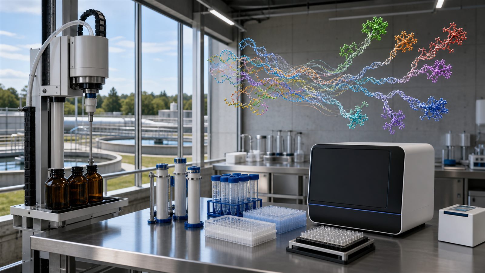
    A vial collected at a wastewater treatment plant can contain biological traces from thousands of people. Public-health laboratories can measure selected pathogens in that mixed sample, and sequencing can reveal some of the genetic variation circulating across the connected community. The result is a population signal that does not depend on everyone seeking care or taking a clinical test.

    That reach makes wastewater surveillance valuable, but it also makes the data easy to misread. A sewer sample is not a census of infections, and a sequence fragment is not a diagnosis. Useful programs combine sampling design, molecular biology, sequencing, bioinformatics, sewer knowledge, clinical surveillance, privacy safeguards, and a clear plan for public-health action.

    The sewer network acts as a pooled sampler

    People with some infections shed viral, bacterial, or other biological material through toilets, sinks, showers, and laundry. Those traces move through sewer pipes and mix before treatment. An autosampler can collect small portions over time to create a composite sample that better represents changing flow than one bottle filled at a single moment.

    The catchment, or area draining to the sampling point, determines what population the result can represent. A sample at a large treatment plant aggregates a broad community. A sample farther upstream can provide more local information, but smaller catchments raise technical, operational, and ethical questions.

    Detection and sequencing answer different questions

    Quantitative polymerase chain reaction, known as qPCR, and digital PCR look for predefined genetic targets. They can sensitively estimate how much target material is present, making them useful for routine trend monitoring. The assay must already know what sequence to search for.

    Sequencing reads many fragments and can help identify lineages, mutations, or multiple targets. Targeted amplicon sequencing amplifies selected genome regions, while hybrid capture uses probes to enrich related sequences. Shotgun metagenomics attempts to read material more broadly without focusing on one pathogen. These approaches trade sensitivity, breadth, cost, speed, and interpretability.

    Wastewater is a difficult molecular mixture

    Pathogen nucleic acids may be rare compared with bacterial, plant, animal, and human material in sewage. Chemicals in the sample can interfere with extraction or amplification. RNA also degrades during travel, storage, and processing. A pathogen that is easy to detect in a clinical swab may be hard to reconstruct from fragmented community wastewater.

    An original comparative study of viral sequencing methods found that untargeted deep sequencing did not provide enough coverage for robust genomic monitoring of low-concentration human viruses in the tested samples. Targeted amplification gave stronger coverage for individual viruses, while hybrid capture offered a way to enrich several viral targets. Method choice must follow the surveillance question.

    A mixed sample contains mixed lineages

    A clinical sequence can often be associated with one infection. Wastewater may contain fragments from many people infected with several lineages. Bioinformatics tools estimate the combination that best explains observed mutations, but fragments do not always show which mutations occurred together in the same genome.

    Low-abundance lineages can be missed, and closely related variants can be hard to separate. Primer-binding changes may cause parts of a genome to drop out during targeted amplification. Results should therefore include uncertainty and quality measures rather than a single overly precise percentage.

    Concentration is not the same as case count

    People shed different amounts of a pathogen at different stages of infection. Sewage flow changes with rainfall, groundwater intrusion, industry, tourism, commuting, and water use. Travel time and temperature affect decay. The number of people connected to a catchment can also change.

    Laboratories may normalize results using flow, population estimates, or biological markers, but no correction removes every source of variation. Trends at one site are usually more reliable than direct comparisons between sites using different collection and laboratory methods. Wastewater can show that a community signal is rising or falling without converting cleanly into a number of infected individuals.

    Timing can provide useful early awareness

    Wastewater does not require a person to recognize symptoms, obtain a test, or report a result. For that reason, a rising signal can sometimes appear before increases become visible in clinical data. The lead time varies with shedding, sampling frequency, laboratory turnaround, healthcare behavior, and the pathogen being monitored.

    CDC describes wastewater monitoring as a complement to other public-health surveillance. That word matters. Hospital admissions, laboratory tests, syndromic reports, genomic sequences from patients, and field investigations provide context that a sewer measurement cannot supply on its own.

    Metadata makes results comparable

    A sequence file without collection context has limited value. Analysts need the date, sampling method, location or catchment, matrix, storage conditions, concentration method, extraction process, sequencing platform, controls, and quality metrics. Standard fields make it easier to combine data and understand why laboratories disagree.

    The US National Center for Biotechnology Information provides a BioSample package for wastewater pathogen genomic surveillance. Shared vocabularies and minimum metadata requirements turn isolated experiments into data that can support national and international analysis.

    Community surveillance requires privacy governance

    Wastewater monitoring is designed to observe pooled biological signals, not identify individuals. Large catchments and fragmented mixed material reduce identifiability, but expanding sequencing capability still deserves governance. Programs should define which targets are justified, how geography is reported, how long raw data is retained, and who can access it.

    Small sites such as buildings, schools, prisons, or workplaces can create greater privacy and stigma concerns than city-scale plants. A technically possible analysis is not automatically appropriate. Public reporting should avoid implying that a neighborhood or institution is responsible for a health threat.

    Multipathogen programs need prioritization

    A sequencing platform can tempt a program to monitor everything detectable. WHO guidance instead emphasizes prioritization and integration with wider surveillance. A target is more useful when it is shed into wastewater, remains detectable, has a validated method, fills an information gap, and can trigger a reasonable response.

    Monitoring many targets also creates quality-control and interpretation burdens. Assays, enrichment panels, reference databases, and reporting rules need maintenance as genomes change. Sustainable staffing and financing matter as much as the sequencer.

    Public dashboards should communicate trends, not certainty

    A dashboard can show relative activity, recent direction, sampling coverage, and confidence. It should also explain missing data, reporting delay, method changes, and the difference between detection and disease burden. A low signal may reflect low circulation, poor recovery, sparse sampling, or a changed assay.

    The same evidence discipline applies to other screening technologies. As our review of multi-cancer blood tests explains, detecting a biological signal does not by itself prove improved outcomes. Surveillance needs a defined decision pathway.

    Sequencing connects biotechnology to public infrastructure

    Wastewater genomics combines treatment plants, cold-chain logistics, automated sample preparation, molecular assays, sequencers, cloud or local computing, and epidemiology. It resembles the broader biotechnology and AI discovery stack, but its output must function as a public service rather than a laboratory demonstration.

    More detailed molecular maps are not always more useful. Spatial proteomics preserves location inside tissue; wastewater deliberately mixes location and people across a catchment. Each technology gains meaning from understanding what its sample represents.

    Limitations

    Coverage is uneven where sewer systems are absent, fragmented, or poorly mapped. Septic systems and informal sanitation may leave populations outside the network. Results can also be distorted by industrial discharges, stormwater, travel, and institutional facilities within a catchment.

    Sequencing cannot prove that a detected organism is alive, infectious, or causing illness. It may not resolve rare lineages or novel pathogens without enough signal and suitable reference methods. Programs should not use community wastewater as a substitute for individual diagnosis or clinical care.

    What to watch next

    Watch for validated multipathogen panels, better recovery controls, standardized metadata, transparent lineage-deconvolution uncertainty, faster reporting, privacy rules for small catchments, and evidence that wastewater signals led to useful actions. WHO’s 2026 landscape analysis also puts governance, ethics, institutional leadership, and sustainable financing at the center of future programs.

    Wastewater sequencing is powerful because it turns a shared infrastructure system into a biological observatory. Its credibility will depend on resisting the urge to turn a mixed community signal into claims the sample cannot support.

    Sources: CDC Wastewater Monitoring Program; CDC information on wastewater data and privacy; WHO 2026 global landscape analysis of wastewater and environmental surveillance; WHO guidance on prioritization, implementation, and integration; Comparison of metagenomic and targeted sequencing methods for wastewater viruses; NCBI BioSample package for wastewater genomic surveillance metadata.

  • Multi-Cancer Blood Tests Need Outcome Evidence, Not Just More Signals

    Multi-Cancer Blood Tests Need Outcome Evidence, Not Just More Signals

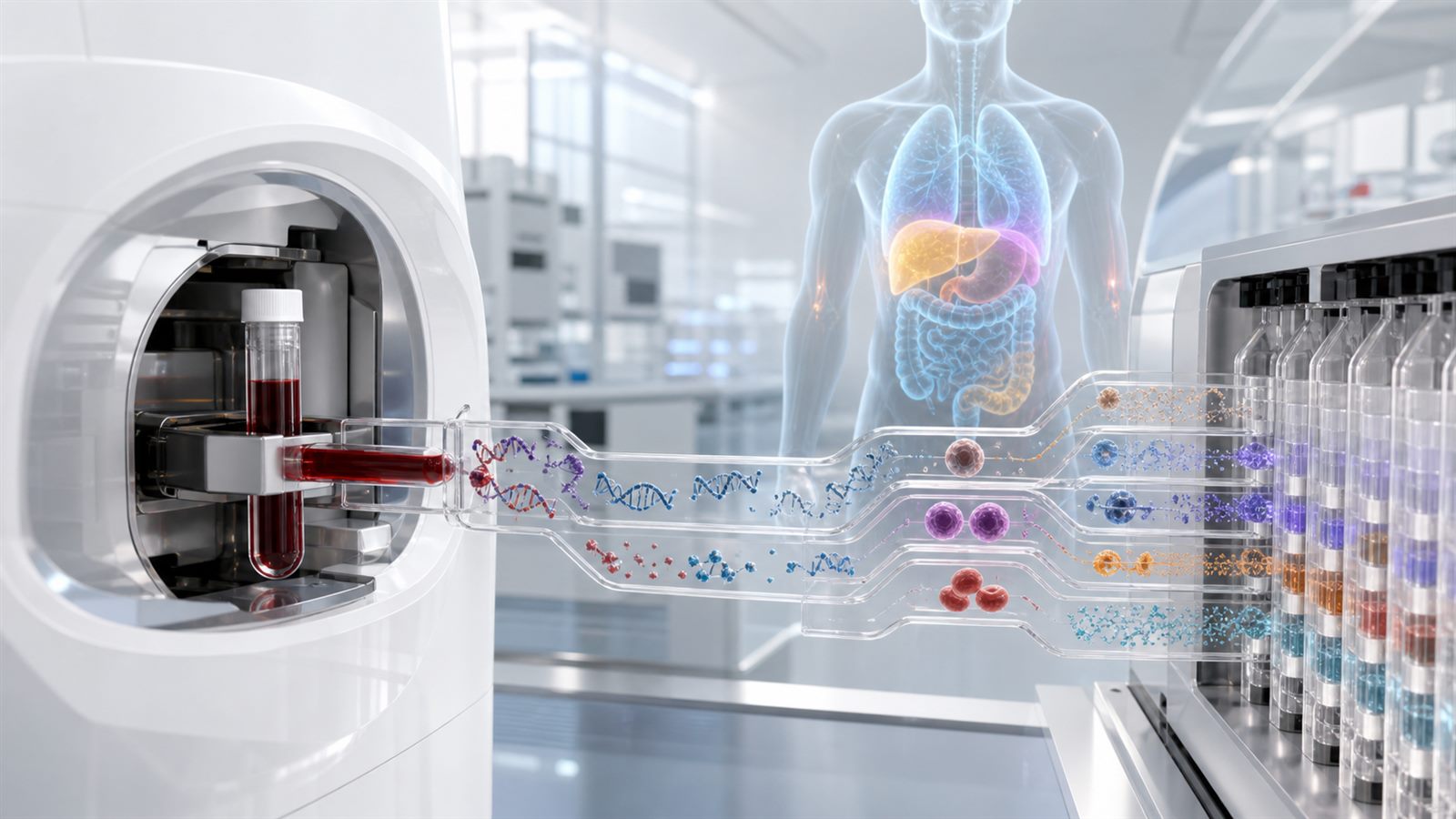
    A single blood draw that looks for many cancers is an appealing idea. Tumors can release fragments of DNA, proteins, cells, and other biological signals into the bloodstream. Multi-cancer detection tests use laboratory instruments and algorithms to search those signals for patterns associated with several cancer types at once.

    The difficult question is not whether a machine can detect a molecular pattern. It is whether screening people without symptoms leads to earlier useful treatment, fewer cancer deaths, and an acceptable burden of false alarms and invasive follow-up. That requires evidence from complete screening pathways, not only impressive laboratory accuracy.

    Multi-cancer tests combine several layers of biotechnology

    A multi-cancer detection test may analyze cell-free DNA, methylation patterns, proteins, circulating tumor cells, or combinations of biomarkers. Sequencing and other high-throughput instruments convert a blood sample into a large set of measurements. A statistical or machine-learning model then estimates whether a cancer-associated signal is present and may predict where in the body it originated.

    This is a different engineering problem from measuring one well-defined substance. The test has to distinguish weak tumor signals from normal biological variation across many people and many possible cancers. Sample collection, storage, laboratory processing, reference data, and model updates can all affect the result.

    Screening is not the same as diagnosis

    A screening test is an initial filter for people who do not have symptoms. A positive result does not by itself establish that cancer is present. It starts a diagnostic process that may include imaging, repeat blood tests, endoscopy, biopsy, or specialist assessment.

    That distinction changes how performance should be judged. The relevant product is not just the assay; it is the assay plus the pathway that resolves positive results. A test that generates a plausible signal but cannot guide doctors toward a timely diagnosis may add anxiety and procedures without delivering a benefit.

    Early-stage sensitivity is the demanding measurement

    Advanced cancers generally release more material into blood than small, localized tumors. The National Cancer Institute notes that current data show multi-cancer tests performing better for later-stage disease than for early-stage disease. Yet the main purpose of population screening is to find important cancers early enough to change an outcome.

    An overall sensitivity number can conceal major differences by cancer type and stage. Readers should look for results broken down by stage, organ, and prespecified population. A test that is very sensitive to a few cancers with strong signals may look impressive in aggregate while missing small tumors that screening most needs to find.

    A tissue-of-origin prediction must be actionable

    When a test reports a possible cancer signal, its predicted tissue of origin helps determine the next examination. A wrong or vague prediction can send the diagnostic search in the wrong direction. Even a correct prediction may not identify a lesion that imaging or biopsy can locate.

    This is why molecular measurement and anatomical confirmation have to work together. Technologies such as spatial proteomics show how much biological context depends on where signals occur inside tissue. A blood test deliberately gives up much of that spatial information for convenience and reach.

    Low prevalence changes the meaning of accuracy

    In a screening population, any one cancer is relatively uncommon. Even a test with high specificity can therefore produce false positives that outnumber true positives. The positive predictive value, meaning the share of positive results that are ultimately confirmed as cancer, depends on both test accuracy and how common the disease is in the tested population.

    Performance from a group already known to contain many cancer cases cannot simply be transferred to average-risk screening. Trials need representative participants without diagnosed cancer and a consistent process for finding which positive and negative results were correct.

    Follow-up burden is part of the safety case

    A useful study should report how many scans, biopsies, specialist visits, and months of uncertainty follow a positive result. It should also record complications and whether a diagnostic search ends without finding cancer. These outcomes matter because screening reaches many healthy people in order to help a much smaller number with previously undetected disease.

    The US Food and Drug Administration has highlighted the complexity of establishing ground truth and designing clinical validation for tests that cover many cancers. Each claimed use, target population, measured analyte, and follow-up pathway affects the balance of probable benefit and harm.

    False negatives and overdiagnosis point in opposite directions

    A false-negative result may create false reassurance. Someone could ignore symptoms or skip established screening because one broad blood test was negative. Multi-cancer testing should therefore be evaluated as an addition to recommended screening, not assumed to replace mammography, cervical screening, colorectal screening, or other proven programs.

    At the other extreme, a test may find a slow-growing cancer that would never have caused illness during a person’s lifetime. That is overdiagnosis. Detecting more cancers is not automatically the same as preventing more deaths, particularly if detection leads to treatment that was not needed.

    Stage shift is useful evidence but not the final outcome

    Researchers may first ask whether screening reduces the number of cancers diagnosed at a late stage. A favorable stage shift would support the idea that tests are finding disease earlier. It is still a surrogate outcome: earlier classification can look better without necessarily extending life, and screening itself can move the recorded diagnosis date forward.

    Randomized trials that measure cancer-specific and overall mortality provide stronger evidence. They take longer and require many participants, but they can capture benefits and harms across the complete care pathway. The same validation discipline applies in other fields, including AI-assisted drug discovery, where generating promising candidates is only the start of proving clinical value.

    NCI is building infrastructure for larger trials

    The National Cancer Institute created the Cancer Screening Research Network to evaluate emerging screening technologies. Its Vanguard Study is a feasibility study for a future, much larger randomized trial of multi-cancer detection. It examines enrollment, test delivery, result return, and how patients and clinicians act after normal or abnormal results.

    That operational work is important. A screening program has to function across laboratories, primary care, imaging centers, and diverse communities. Evidence from a tightly controlled research cohort may not predict access, adherence, or diagnostic delays in everyday care.

    What evidence should technology readers ask for?

    Useful reports should separate analytical validity, clinical validity, and clinical utility. Analytical validity asks whether the laboratory measures its target reliably. Clinical validity asks how accurately the result identifies cancer in the intended population. Clinical utility asks whether using the result improves health outcomes enough to justify the harms and cost.

    Readers should also check whether the validation population is independent of the data used to train the algorithm, whether results are reported for early stages, and whether performance is consistent across age, sex, ancestry, health conditions, and collection sites. As with tumor sequencing for personalized vaccines, the molecular test is only one part of a much larger clinical and manufacturing system.

    Limitations

    Multi-cancer tests vary in biomarkers, algorithms, target populations, and claimed cancer coverage, so one result cannot represent the whole category. Commercial availability does not establish that a test reduces mortality. Regulatory status and clinical evidence can also change, and screening decisions belong in a conversation with a qualified health professional.

    This article is a technology overview, not personal medical guidance. A broad blood test should not be treated as a substitute for symptoms being assessed or for established screening recommended for an individual’s age and risk.

    What to watch next

    Watch for randomized evidence on late-stage incidence and mortality, standardized diagnostic pathways after a positive signal, detailed reporting of false positives and unresolved cases, and results from populations that reflect real screening programs. Better assays will matter, but so will faster imaging, reliable referrals, and equitable access to follow-up.

    Multi-cancer detection may eventually expand the reach of screening. The decisive milestone will not be finding more molecular signals in blood; it will be proving that acting on those signals helps people more than it harms them.

    Sources: National Cancer Institute questions and answers on multi-cancer detection tests; NCI Vanguard Study record; NCI Cancer Screening Overview; FDA advisory meeting materials on multi-cancer detection tests.

  • Xenotransplantation Is Becoming a Clinical Trial and Surveillance System

    Xenotransplantation Is Becoming a Clinical Trial and Surveillance System

    Transplanting a genetically engineered pig organ into a person is moving from isolated experimental cases toward structured clinical research. That is a major scientific transition, but it is not the same as proving that xenotransplantation is ready for routine care. A regulated trial must show that an organ can function, that immune rejection can be controlled, and that risks can be monitored over time.

    The distinction matters because xenotransplantation is not just a surgical procedure. It is a tightly connected biotechnology system involving the donor animal, genetic edits, pathogen screening, organ preservation, immunosuppressive treatment, recipient follow-up, and public-health surveillance. The clinical result depends on every part of that chain.

    Why researchers use genetically engineered pigs

    Pig organs are similar enough in size and function to make them plausible candidates for human transplantation, and pigs can be bred under controlled conditions. The biological barrier is severe, however. Unmodified pig cells display molecules that the human immune system can recognize as foreign, creating a risk of rapid rejection. Differences in complement regulation, blood coagulation, inflammation, and organ growth can create additional problems.

    Gene editing lets developers remove selected pig genes and add selected human genes intended to make the organ more compatible. This does not make the organ human, and a larger number of edits is not automatically better. Each combination needs evidence showing what a change does, whether it creates unintended effects, and whether the engineered trait remains stable across donor animals.

    A clinical trial changes the evidence standard

    Earlier living-recipient procedures often occurred through special regulatory pathways for patients with serious conditions and limited alternatives. Those cases provided valuable observations, but different patients, products, and treatment protocols make broad conclusions difficult.

    ClinicalTrials.gov currently lists NCT06878560, an interventional Phase I/II study of a ten-gene-edited pig kidney in people with end-stage renal disease. The registry lists a planned enrollment of 50 and describes the study as recruiting. Those facts establish that a formal protocol exists; they do not establish safety or effectiveness. Early-stage trials are designed to generate that evidence under predefined eligibility, monitoring, and reporting rules.

    The donor animal is part of the medical product

    For a conventional manufactured medicine, quality teams test ingredients, process controls, and finished batches. Xenotransplantation adds a living source animal. Regulators need information about the breeding herd, its genetic lineage, housing, feed, health records, and exposure to infectious agents. The organ’s identity must remain traceable from the donor animal to the recipient.

    Consistency is difficult because biology varies. A program must show that intended genetic edits are present, that unexpected edits or mosaicism are controlled, and that organ handling follows a repeatable process. This platform challenge resembles the evidence-reuse question in gene-editing therapies: shared technology can support common knowledge, but each final product still needs its own proof.

    Immune rejection remains a moving target

    Removing major incompatibility signals may prevent one form of immediate rejection, but the immune system has many pathways. Antibodies, complement proteins, immune cells, coagulation factors, and inflammation can damage a graft over different timescales. Recipients also need immunosuppressive treatment, which can introduce infection and other complications.

    Researchers therefore track more than whether the organ produces urine or filters blood on a particular day. They monitor kidney function, tissue injury, antibody responses, clotting, medication levels, imaging, biopsies, and adverse events. A useful trial must connect clinical changes to both the engineered organ and the treatment regimen around it.

    Infectious-disease surveillance extends beyond the recipient

    Moving living animal cells into a person creates a theoretical route for animal pathogens to adapt or spread. Controlled herds and extensive screening reduce that risk but cannot turn it into zero. Some organisms may be difficult to detect, and an infection could emerge after the transplant.

    FDA and U.S. public-health guidance therefore treats infectious-disease monitoring as a central part of xenotransplantation. Programs need archived samples, validated assays, recipient surveillance, traceability, and plans for investigating unexpected illness. A published report on the first living recipient of a genetically modified pig kidney described donor screening, pathogen mitigation, and post-transplant monitoring as an integrated strategy rather than a one-time test.

    Trials must measure function and durability

    An organ can begin working and still fail later. Trials need to examine the duration and quality of graft function, rejection episodes, complications from immunosuppression, hospital use, patient survival, and quality of life. They also need clear rules for removing a failing graft or returning a patient to dialysis where possible.

    Small early cohorts can identify major safety signals and improve protocols, but they cannot reveal every uncommon event or predict long-term performance. The same reproducibility discipline discussed for organoid biotechnology platforms applies here at a much higher clinical stake: measurements, definitions, and sample handling must be consistent enough for results to be compared.

    Consent has unusual long-term obligations

    A participant must understand that the organ is experimental, that outcomes are uncertain, and that follow-up may be intensive. Monitoring requirements may continue even if the person feels well. Family members and close contacts may also need information if investigators suspect an infectious risk.

    Privacy and public health can pull in different directions. Researchers need enough traceability to investigate transmission while protecting medical and genetic information. Trial design should explain who holds samples and data, how long they are retained, what findings will be returned, and what happens if a participant later wants to withdraw.

    Scaling requires more than successful surgery

    Even if trials show benefit, a clinical service would need specialized breeding facilities, standardized editing, reliable organ recovery, transport logistics, trained transplant teams, pathogen laboratories, and long-term registries. Costs and access will depend on whether those systems can operate consistently across centers.

    Data integration will be crucial. Tissue measurements such as those described in spatial proteomics may help researchers understand where rejection and inflammation occur, but exploratory biomarkers must be linked to validated clinical outcomes.

    Limitations

    Current evidence includes small numbers of living recipients, nonhuman-primate studies, decedent studies, and trials with products that do not all use the same genetic edits or immunosuppressive protocols. Outcomes from one organ type cannot simply be transferred to another. Public trial registries describe planned methods, not completed results.

    Xenotransplantation also raises animal-welfare, allocation, consent, and equity questions that laboratory performance cannot resolve alone. Oversight will need medical, public-health, ethical, and patient perspectives.

    What to watch next

    Watch for peer-reviewed trial results that report graft function, rejection, infections, adverse events, and protocol changes rather than only procedure announcements. Also watch whether different centers can reproduce donor screening and clinical monitoring, and whether regulators update guidance as evidence accumulates.

    The real milestone is not one successful operation. It is a transparent clinical and surveillance system that can determine who benefits, for how long, and at what risk.

    Sources: FDA xenotransplantation guidance; ClinicalTrials.gov: NCT06878560; American Journal of Transplantation: Infectious disease surveillance in clinical xenotransplantation; Nature Communications study of genetically engineered pig kidney grafts.

  • Spatial Proteomics Is Turning Tissue Slides Into Molecular Maps

    Spatial Proteomics Is Turning Tissue Slides Into Molecular Maps

    Biology is becoming more spatial. For decades, many laboratory tests treated tissue as if it were a blended sample: grind it up, measure molecules, and compare the totals. That approach can be powerful, but it loses an important fact about living systems. Cells do not act alone. They sit next to other cells, receive local signals, form structures, and change behavior depending on where they are in a tissue.

    Spatial proteomics tries to keep that context. Instead of asking only which proteins are present, it asks where proteins are located, which cell neighborhoods they appear in, and how those patterns change across healthy tissue, disease, treatment, or development. That makes the field important for biotech because proteins are often closer to biological function than genes alone. They are the machines, receptors, enzymes, markers, and signals that drugs frequently target.

    What spatial proteomics measures

    Proteomics is the large-scale study of proteins. Spatial proteomics adds location. A tissue section can be stained with many antibodies, imaged in repeated cycles, or analyzed with mass-spectrometry methods that preserve positional information. The output is not just a table. It is closer to a molecular map, where protein patterns can be tied back to tissue architecture.

    Nature Methods named spatial proteomics its 2024 Method of the Year, highlighting how these methods are helping atlas-scale projects understand biological complexity in health and disease. That recognition is useful because the field is not one single machine or assay. It includes microscopy-heavy approaches, mass-spectrometry approaches, and hybrid pipelines that combine imaging, segmentation, data integration, and statistical modeling.

    Why tissue maps matter

    The Human BioMolecular Atlas Program, or HuBMAP, is one of the clearest examples of where this is going. The HuBMAP Consortium is building tools and datasets for mapping the healthy human body across cells, molecules, and organs. Its goal is not merely to collect attractive images. It is to build reference maps that help researchers compare healthy tissue with aging, disease, injury, and treatment response.

    Reference maps can also reduce confusion. If a protein signal appears in a cancer biopsy, researchers need to know whether that pattern is unusual or part of normal tissue variation. If a drug changes immune-cell neighborhoods, scientists need to know whether the shift is linked to benefit, toxicity, or neither. Spatial maps provide a richer baseline than bulk measurements alone.

    They can also help bridge discovery and translation. A biomarker that looks promising in a small study is more convincing when researchers can show where it appears, which neighboring cells surround it, and whether the pattern is consistent across independent tissue collections.

    Proteins add practical biology

    Single-cell RNA sequencing has transformed biology by showing which genes are active in individual cells. But RNA is not the whole story. A cell may transcribe a gene without producing much protein, and protein location inside or around the cell can change function. The Human Protein Atlas is valuable partly because it connects protein expression and localization across tissues, cells, subcellular compartments, blood, and disease contexts.

    For ordinary technology readers, this is the key point: spatial proteomics is a data infrastructure technology for biology. It helps turn slides into searchable, comparable maps. That makes it relevant to the same biotech platform questions we covered in organoid reproducibility and gene-editing evidence reuse. Better maps do not automatically make better therapies, but they can make experiments more interpretable.

    Where it could help first

    Cancer research is an obvious use case because tumors are spatially messy. Cancer cells, immune cells, blood vessels, fibroblasts, necrotic regions, and treatment-resistant pockets can sit next to one another. A bulk sample may say that a marker is present. A spatial proteomics experiment can show whether that marker is on the tumor edge, inside an immune-rich region, or concentrated around blood vessels.

    Inflammation, neuroscience, kidney disease, placental biology, and drug-toxicity research are also natural fits. In each case, location matters. A protein signal in the wrong cell type or tissue layer can mean something very different from the same signal elsewhere. This is why spatial analysis can complement manufacturing-style biotech workflows such as personalized cancer vaccines, where tumor context may affect which targets are meaningful.

    The limits are real

    Spatial proteomics is not a magic microscope. Experiments can be expensive, technically demanding, and hard to compare across laboratories. Antibodies vary in quality. Tissue preparation can change signals. Imaging cycles can introduce artifacts. Segmentation software may misidentify cell boundaries, especially in dense tissue. Different platforms may measure different panels of proteins at different resolutions.

    Data analysis is another bottleneck. A single tissue section can produce large images, thousands to millions of cells, and many protein channels. Researchers need metadata, quality controls, coordinates, image-processing pipelines, and statistical methods that preserve biological meaning. A beautiful tissue map is less useful if other teams cannot reproduce, query, or combine it with related datasets.

    What to watch next

    Watch for more standardized panels, better public datasets, and tools that connect spatial proteomics with genomics, transcriptomics, clinical records, and drug-response data. Also watch how atlas projects handle consent, privacy, tissue provenance, and representation across populations. A reference atlas is only as useful as the quality and diversity of the data behind it.

    The practical promise is not instant personalized medicine. It is a better way to describe the physical organization of biology. If biotech is going to design more precise therapies, it needs to know not only which molecules exist, but where they act. Spatial proteomics gives researchers a sharper map of that terrain.

    Sources: HuBMAP Consortium; Human Protein Atlas; Nature Methods: Method of the Year 2024, spatial proteomics.

  • Organoids Need Reproducibility Before They Can Become Routine Biotech Platforms

    Organoids Need Reproducibility Before They Can Become Routine Biotech Platforms

    Organoids are miniature, simplified tissue models grown from cells. They can mimic selected features of organs such as the intestine, brain, liver, or lung, and they are becoming important tools for drug development, disease research, and personalized medicine. But the excitement around organoids can hide a hard engineering problem: reproducibility.

    If two laboratories grow organoids from similar cells and get different results, the model may still be scientifically interesting, but it becomes harder to use for routine decisions. For organoids to move from promising research tools to dependable biotechnology platforms, scientists need better standards for cell sourcing, culture conditions, measurements, and data reporting.

    Organoids Are Powerful Because They Preserve Context

    Traditional cell cultures often grow as flat layers. They are useful, but they can miss three-dimensional structure, cell diversity, and tissue-like behavior. Organoids can preserve more of that context, which is why researchers use them to study development, disease mechanisms, infection, toxicity, and drug response.

    The National Institutes of Health supports this direction through programs such as the Standardized Organoid Modeling program. Its focus on reproducible organoid models is a signal that the field is moving beyond one-off demonstrations. The goal is not simply to make organoids, but to make organoids that other researchers can understand and compare.

    This builds on the broader shift we covered in organs-on-chips and regulatory tools. Better human-relevant models can reduce dependence on less predictive systems, but only if the model itself is well characterized.

    Reproducibility Starts With Cell Identity

    An organoid is not a generic product. Its behavior depends on the starting cells, donor background, genetic changes, reprogramming methods, passage number, and culture history. Two models called by the same tissue name may not be equivalent.

    That makes cell identity and provenance essential. Researchers need to document where cells came from, how they were handled, what quality checks were performed, and whether the cells changed over time. Without those details, a surprising result may reflect biology, contamination, drift, or a subtle difference in protocol.

    The same issue appears in genome-editing safety analysis. Biology is sensitive to starting conditions, and good measurement begins with knowing exactly what system is being measured.

    Culture Conditions Can Change the Outcome

    Organoids depend on media, growth factors, extracellular matrix materials, oxygen levels, timing, mechanical conditions, and handling technique. Small changes can influence size, maturation, cell composition, and response to drugs.

    That is a problem for scale. A model that works in one expert lab may not transfer cleanly to a contract research organization, pharmaceutical screening facility, or hospital-linked testing program. Automation can help, but automation must be paired with validated protocols and quality controls.

    Useful standards should describe not only the recipe, but the acceptable range of variation. If organoids are too large, too immature, too heterogeneous, or missing key cell types, the downstream assay may look precise while measuring the wrong thing.

    Measurements Need Reference Points

    Researchers often measure organoids using imaging, sequencing, protein markers, functional assays, and drug-response curves. Each method adds information, but each method also creates choices. Which markers define maturity? Which imaging features indicate healthy structure? Which statistical threshold separates a true drug effect from noise?

    Reference data can help. A well-described organoid model should be compared with relevant human tissue, known controls, and expected failure modes. It should also include uncertainty. A clean chart without error estimates can be misleading.

    The US Food and Drug Administration’s work on advancing alternative methods shows why evidence quality matters. New approach methodologies can be valuable, but regulators need to understand when a model is fit for a particular purpose.

    Drug Screening Raises the Stakes

    Organoid drug screening is attractive because it may reveal human-relevant responses earlier than some traditional tests. Cancer organoids, for example, can sometimes be grown from patient tumor samples and exposed to candidate therapies. Disease organoids can help compare compounds in a more tissue-like setting.

    But screening is only useful when the assay is stable. If organoid size, composition, or maturity varies across plates, drug-response differences may come from manufacturing noise. High-throughput screening also needs consistent imaging, dosing, incubation, and analysis pipelines.

    This does not make organoids unreliable. It means the field is entering a manufacturing and quality-systems phase, similar to the platform questions in gene-editing therapy development. Powerful biology needs disciplined process control.

    What Better Reporting Should Include

    • Cell source, donor information where appropriate, passage number, and authentication method.
    • Culture media, matrix, growth factors, timing, oxygen conditions, and handling steps.
    • Quality-control metrics for size, morphology, marker expression, viability, and contamination.
    • Assay protocols, controls, analysis software, and uncertainty estimates.
    • Known limitations, failed batches, and conditions where the model should not be used.

    These details can feel tedious, but they decide whether other labs can reproduce a result. They also help companies decide whether an organoid model is ready for screening, validation, or regulatory discussion.

    What to Watch Next

    Watch for more public reference organoid models, shared protocols, benchmark datasets, and regulatory case studies that define fit-for-purpose use. The most important progress may not look like a dramatic image. It may look like boring metadata, quality criteria, and documented failure modes.

    Organoids can make biotechnology more human-relevant, but they will earn trust through repeatability. The next phase is less about proving that miniature tissues can grow and more about proving that they can be measured, compared, and used responsibly.

    Sources and Further Reading