
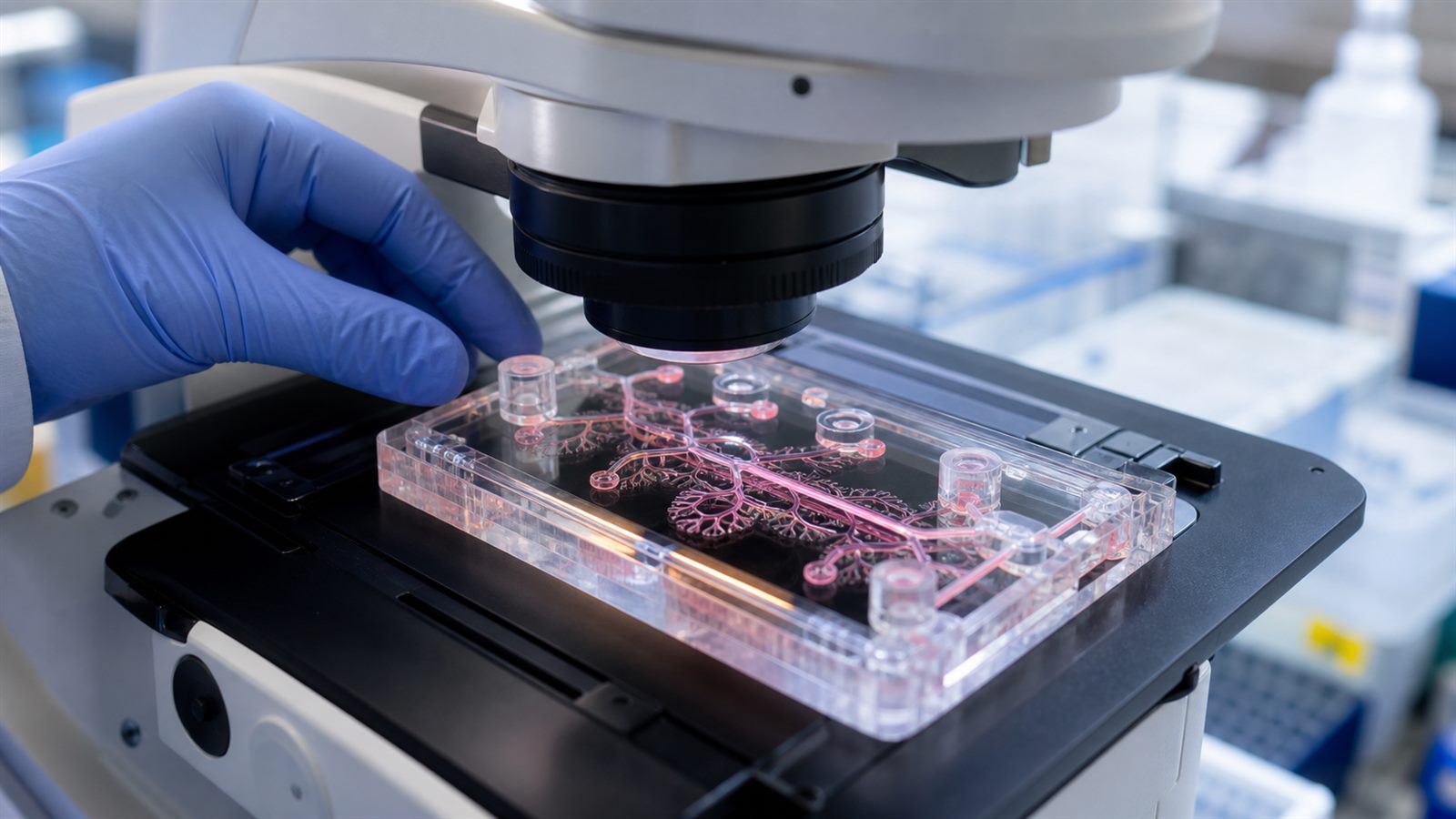
An organ-on-a-chip is not a miniature replacement organ. It is a small engineered device that places living human cells in controlled channels, chambers, membranes, and fluid flows so researchers can reproduce selected features of an organ. A lung model might expose cells to air on one side and flowing medium on the other. A liver model may be designed to study metabolism and toxicity. A blood-brain barrier model focuses on how substances cross a protective interface.
These systems, also called tissue chips or microphysiological systems, are moving from experimental curiosities toward drug-development tools. The shift is being driven by better cell biology, microfluidics, measurement, and a regulatory push for validated human-relevant testing methods. Their promise is substantial, but their value depends on defining exactly what each model can predict.
What a Tissue Chip Recreates
Traditional cell culture often grows a single cell type on a flat plastic surface. A tissue chip can add three-dimensional structure, multiple cell types, mechanical forces, chemical gradients, and continuous flow. Those features can influence how cells mature, communicate, and respond to a drug.
The chip does not need to copy every feature of an organ to be useful. It may model one decision-relevant function: whether a compound damages liver cells, crosses a barrier, triggers inflammation, or changes the rhythm of heart tissue. The intended purpose is called the context of use, and it determines what evidence the model needs.
Organoids are related but not identical. They are three-dimensional cell structures that can self-organize into features resembling a tissue. Organ-on-chip systems add an engineered environment around cells and may incorporate an organoid. Both belong to the wider set of new approach methodologies, or NAMs, alongside computational models, biochemical assays, and other non-animal methods.
Why Drug Development Needs Better Models
A drug candidate can look promising in a simple assay and still fail because the human body absorbs, metabolizes, or reacts to it differently. Animal studies provide whole-organism information, but species differences can limit their ability to predict a specific human response. Clinical trials remain essential, yet they occur after years of laboratory work and cannot be the first place every avoidable toxicity is discovered.
Human-cell models can add evidence earlier. Researchers may compare healthy and diseased tissue, test several concentrations, or use cells with a particular genetic background. This can help prioritize candidates and investigate why a response occurs. It complements the validation problem described in our article on AI-assisted drug discovery: generating candidates faster is useful only when experiments can reliably reject weak ideas.
FDA Guidance Is Raising the Validation Bar
The US Food and Drug Administration’s 2025 roadmap promoted human-based lab models, organ-on-chip systems, cell assays, and computational approaches as alternatives that may reduce or replace some animal testing. In March 2026, the agency released draft guidance describing general considerations for validating NAMs submitted in drug development.
The guidance highlights four principles. First, the context of use must be clear. Second, the model should have relevant human biology for the question. Third, its technical performance should be robust, reliable, and reproducible. Fourth, it must be fit for the regulatory decision it is meant to support.
Those principles prevent a common misunderstanding. A visually impressive chip is not automatically a validated predictor. Developers need evidence about cell sources, device materials, fluid conditions, measurements, controls, variability, and performance across laboratories. The required evidence changes with the consequence of a wrong answer.
Reproducibility Is the Engineering Challenge
Living systems vary. Cells from different donors may behave differently, and cells produced from induced pluripotent stem cells may mature unevenly. Tiny changes in channel geometry, coating, flow, oxygen, temperature, or sampling time can alter results. Some device materials absorb drug molecules, changing the concentration that cells actually receive.
A useful platform therefore needs quality controls and reference compounds. Researchers should know whether a failed run came from the drug, the cells, the chip, or the measurement. Standard operating procedures and shared data formats are as important as the microfluidic hardware.
The National Center for Advancing Translational Sciences coordinates tissue-chip research with other NIH institutes and the FDA. Its Translational Centers for Microphysiological Systems are intended to strengthen models and help them meet the requirements for qualified drug-development tools. Cross-laboratory testing is critical because a result that works only in the inventor’s lab is difficult to use in a regulatory submission.
One Chip Cannot Represent the Whole Body
A drug may be absorbed in the gut, transformed in the liver, cleared by the kidneys, and affect the heart. Linking several tissue models could reveal interactions that a single chip misses. Researchers are developing multi-organ systems with controlled fluid exchange, but integration adds complexity. The relative size of each tissue, fluid volume, timing, and common culture medium all influence whether the combined model remains biologically meaningful.
Immune responses, hormones, nerves, microbiomes, aging, and chronic exposure can also be difficult to reproduce. Some adverse effects are rare or emerge only after long use. Tissue chips should therefore be viewed as one evidence layer within a broader testing strategy, not as a universal replacement for every laboratory method, animal study, or clinical trial.
Patient-Specific Models Have a Different Promise
Cells derived from a patient can, in principle, help researchers study a rare disease or compare possible treatments for a biological subtype. That does not mean a chip can currently choose the right medicine for an individual with certainty. Manufacturing time, cell maturity, cost, and clinical validation remain substantial barriers.
The careful, one-patient development described in our article on personalized CRISPR therapy shows why customized models are attractive and why evidence standards cannot be relaxed. A patient-specific chip may generate a useful hypothesis, but treatment decisions still require clinical oversight and validated interpretation.
What the Public Should Not Infer
A regulator accepting NAM data does not mean every product can skip animal studies or proceed directly to people. The FDA draft guidance is general and tells developers to consult the appropriate review division for the specific disease, organ, endpoint, and product. Acceptance depends on context and evidence.
Claims that a chip will make drug development automatically faster or cheaper should also be treated cautiously. Better early prediction could reduce wasted work, but building, validating, and operating sophisticated human-cell systems requires investment and specialist expertise. Benefits will appear first where a model answers a defined question better than the current method.
What to Watch Next
Watch for qualified drug-development tools, studies reproduced across independent laboratories, reference data sets, and regulatory submissions that explain how chip results changed a decision. Progress will also depend on reliable cell production, sensors that can monitor tissue without destroying it, and agreed ways to combine chip data with computational models.
Tissue chips sit at the intersection of the programmable biology discussed in our synthetic biology overview and the practical demands of drug safety. Their success will not be measured by how closely a device looks like an organ. It will be measured by whether it produces a reproducible answer that helps protect people.
Leave a Reply